Beranda
/ Lower Body Diagram - Lower Limb Bones Illustrations Radiology Case Radiopaedia Org - Did you know… ☛ while the size of the human head right from birth won't change drastically, it is the torso and the lower limbs that grow in length.
Lower Body Diagram - Lower Limb Bones Illustrations Radiology Case Radiopaedia Org - Did you know… ☛ while the size of the human head right from birth won't change drastically, it is the torso and the lower limbs that grow in length.
Insurance Gas/Electricity Loans Mortgage Attorney Lawyer Donate Conference Call Degree Credit Treatment Software Classes Recovery Trading Rehab Hosting Transfer Cord Blood Claim compensation mesothelioma mesothelioma attorney Houston car accident lawyer moreno valley can you sue a doctor for wrong diagnosis doctorate in security top online doctoral programs in business educational leadership doctoral programs online car accident doctor atlanta car accident doctor atlanta accident attorney rancho Cucamonga truck accident attorney san Antonio ONLINE BUSINESS DEGREE PROGRAMS ACCREDITED online accredited psychology degree masters degree in human resources online public administration masters degree online bitcoin merchant account bitcoin merchant services compare car insurance auto insurance troy mi seo explanation digital marketing degree floridaseo company fitness showrooms stamfordct how to work more efficiently seowordpress tips meaning of seo what is an seo what does an seo do what seo stands for best seotips google seo advice seo steps, The secure cloud-based platform for smart service delivery. Safelink is used by legal, professional and financial services to protect sensitive information, accelerate business processes and increase productivity. Use Safelink to collaborate securely with clients, colleagues and external parties. Safelink has a menu of workspace types with advanced features for dispute resolution, running deals and customised client portal creation. All data is encrypted (at rest and in transit and you retain your own encryption keys. Our titan security framework ensures your data is secure and you even have the option to choose your own data location from Channel Islands, London (UK), Dublin (EU), Australia.
Lower Body Diagram - Lower Limb Bones Illustrations Radiology Case Radiopaedia Org - Did you know… ☛ while the size of the human head right from birth won't change drastically, it is the torso and the lower limbs that grow in length.. The spine anatomy is a complex structure. The vertebrae, which stack like spools of thread, support the back and protect the spinal cord. Evenly distribute weights from your upper body into the lower extremities In addition to bearing the weight of the upper body, the knee allows for walking, running, and jumping. The spine diagram shown below, consists of many bones or vertebrae,soft discs,the spinal cord, and spinal nerves.
We hope this picture human body artery diagram in detail can help you study and research. Arteries (in red) are the blood vessels that deliver blood to the body. By definition, an artery is a vessel that conducts blood from the heart to the periphery. Quadriceps (made of 4 muscles): • removing the pelvic lymph nodes • removing the groin lymph nodes • having pelvic and/or groin radiation this pamphlet explains:
6 Best Leg Exercises For Mass Barbend Leg Muscles Anatomy Leg Muscles Diagram Hip Muscles Anatomy from i.pinimg.com Stronger muscles can help stabilize the lower back and can help reduce injury risk. Key bones in the abdominal area include the base of the ribcage and the lumbar spine in the lower back. Health conditions associated with the lower respiratory system. The iliac, femoral, popliteal and tibial (calf) veins are the deep veins in the legs. At the level of the pelvic bones, the abdomen. Muscle anatomy gluteus 12 photos of the muscle anatomy gluteus gluteus muscle anatomy ct, gluteus muscle anatomy mri, human muscle anatomy gluteus maximus, muscle anatomy gluteus, muscle anatomy of gluteal, human muscles, gluteus muscle anatomy ct, gluteus muscle anatomy mri, human muscle anatomy. This section of the nervous system features the most inferior portion of the spinal cord along with many major nerves, plexuses, and ganglia that serve the vital organs of the. Balance the weight of your head on top of your spine;
Anatomynote.com found human body artery diagram in detail from plenty of anatomical pictures on the internet.
1 your spine in this region has a natural inward curve. The torso of the human body also consists of the major muscles of our body; 2) the tension force t ' 3 exerted by the string on the block. Lower respiratory tract infection (lrti) pneumonia Ascending aorta, aortic arch, thoracic aorta, and abdominal aorta. Proper posture and body mechanics. By definition, an artery is a vessel that conducts blood from the heart to the periphery. A) free body diagram for the block; Posted on may 24, 2016 by admin. We think this is the most useful anatomy picture that you need. Superficial and deep anterior muscles of upper body Learn vocabulary, terms, and more with flashcards, games, and other study tools. This curve, called lordosis, helps to:
Deep veins, located in the center of the leg near the leg bones, are enclosed by muscle. Learn vocabulary, terms, and more with flashcards, games, and other study tools. Key bones in the abdominal area include the base of the ribcage and the lumbar spine in the lower back. Muscle anatomy gluteus 12 photos of the muscle anatomy gluteus gluteus muscle anatomy ct, gluteus muscle anatomy mri, human muscle anatomy gluteus maximus, muscle anatomy gluteus, muscle anatomy of gluteal, human muscles, gluteus muscle anatomy ct, gluteus muscle anatomy mri, human muscle anatomy. Below you'll see diagrams along with the names of the back muscles that may be the cause of your pain.
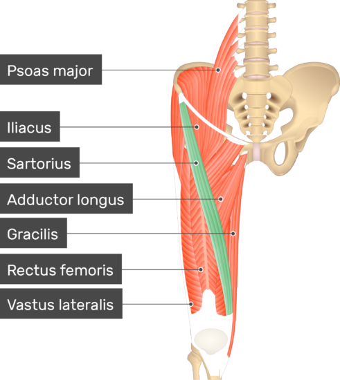
Sartorius Muscle from www.getbodysmart.com Your lower back (lumbar spine) is the anatomic region between your lowest rib and the upper part of the buttock. Did you know… ☛ while the size of the human head right from birth won't change drastically, it is the torso and the lower limbs that grow in length. Related posts of muscles of the lower back and buttocks diagram muscle anatomy gluteus. At the level of the pelvic bones, the abdomen. The spine diagram shown below, consists of many bones or vertebrae,soft discs,the spinal cord, and spinal nerves. Labeled illustration chart on white. It's also the largest joint in the body. Likewise, there are muscles in other parts of the body that help support and move the spine.
Lower thoracic, lumbar vertebrae and sacrum:
Related posts of muscles of the lower back and buttocks diagram muscle anatomy gluteus. Evenly distribute weights from your upper body into the lower extremities In addition to bearing the weight of the upper body, the knee allows for walking, running, and jumping. Deep veins, located in the center of the leg near the leg bones, are enclosed by muscle. Veins (in blue) are the blood vessels that return blood to the heart. Posted on may 24, 2016 by admin. The torso of the human body also consists of the major muscles of our body; It contains partially digested food before it. Superficial and deep anterior muscles of upper body Key bones in the abdominal area include the base of the ribcage and the lumbar spine in the lower back. This article looks at female body parts and their functions, and it provides an interactive diagram. Anatomynote.com found human body artery diagram in detail from plenty of anatomical pictures on the internet. Stronger muscles can help stabilize the lower back and can help reduce injury risk.
How many muscles are in the back? This is the lower part of the stomach. Related posts of muscles of the lower back and buttocks diagram muscle anatomy gluteus. Deep veins, located in the center of the leg near the leg bones, are enclosed by muscle. By definition, an artery is a vessel that conducts blood from the heart to the periphery.
Anatomy Of The Human Body Information Infographic Free Vector On Freepik from img.freepik.com The vertebrae, which stack like spools of thread, support the back and protect the spinal cord. Labeled illustration chart on white. Lower thoracic, lumbar vertebrae and sacrum: Stronger muscles can help stabilize the lower back and can help reduce injury risk. Learn vocabulary, terms, and more with flashcards, games, and other study tools. It contains partially digested food before it. The knee joins the upper leg and the lower leg. If you are using a lower power led, you will need to use longer treatment times, and proportionally less joules.
The spine diagram shown below, consists of many bones or vertebrae,soft discs,the spinal cord, and spinal nerves.
Your lower back (lumbar spine) is the anatomic region between your lowest rib and the upper part of the buttock. Woman holding a blackboard with an illustration of the human digestive system drawn on it in chalk. Stronger muscles can help stabilize the lower back and can help reduce injury risk. 1 your spine in this region has a natural inward curve. Anatomynote.com found human body artery diagram in detail from plenty of anatomical pictures on the internet. This article looks at female body parts and their functions, and it provides an interactive diagram. Ascending aorta, aortic arch, thoracic aorta, and abdominal aorta. Evenly distribute weights from your upper body into the lower extremities Moves humerus (arm) to chest. Muscle anatomy gluteus 12 photos of the muscle anatomy gluteus gluteus muscle anatomy ct, gluteus muscle anatomy mri, human muscle anatomy gluteus maximus, muscle anatomy gluteus, muscle anatomy of gluteal, human muscles, gluteus muscle anatomy ct, gluteus muscle anatomy mri, human muscle anatomy. Superficial and deep anterior muscles of upper body B) free body diagram of point p; The pectoral muscles, the abdominal muscles, and the lateral muscle.